Arterial Assisted Stretching
For Healing Scoliosis
See Real Spine Transformations
Watch verified pre & post-treatment videos of crooked spines becoming straight during hands-on sessions with Dr. Clay and the practitioners he has trained.
View VideosEmpirical
Impeccable
Unimpeachable
Developed by
Dr. Cassius Camden Clay, Chiropractor
As of January 1, 2026, of the 70 people whom I have treated for scoliosis over the past six months, 69 completely resolved with a 5 to 10 minute treatment, mostly by gently tugging on the carotid arteries.
69 crooked spines became perfectly straight!
One in five people, whom I have examined, have mild to moderate scoliosis.
Most scolioses exist in the middle back.
Be Realistic, Expect a Miracle!
I learned an osteopathic technique in June of 2025, where I witnessed restricted neck motions of right rotation and right lateral leaning, which resolved by releasing tension in the left common carotid artery found in the front of the neck.
Carotid artery relaxation allowed return of the neck’s full ranges of motion.
A gentle tug for a very short period of time on the left carotid artery—pulling away from the brain and toward the heart—released tension in the arteries in the brain, thereby relaxing the left carotid artery. Once this artery was relaxed, tension and pain in the neck were completely resolved.
WOW!
One of my discoveries and specialties for the past 15 years is
“Scoliosis Resolution.”
The basic concept is the practitioner tugs the vertebra at the apex of the scoliosis counterintuitively to make it worse. The body pulls the spine at the apex against the practitioner, pulling the spine straight—usually in less than two minutes.
This works almost every time to obliterate mild to moderate scoliosis in the middle back, resulting in a straight spine and the comfort that comes with it.
A video and book on this technique may be found at
www.QuickSelfFixes.com
.
Go to “Free Videos & Books.”
There you will find “Scoliosis Resolution” (video & book) and
“Self-Scoliosis Resolution” (video) near the top of the list.
From the osteopathic technique for correcting restrictions of neck ranges of motion, I applied this concept to correcting spinal scoliosis.
“Arterial Assisted Stretching” causes arterial relaxation in the arteries, focusing on the arteries in the heart muscle.
In most cases, within 5 to 10 minutes, “Arterial Assisted Stretching” will straighten a spine from mild to moderate scoliosis in the middle back.
The concept which releases reduced neck ranges of motion also heals scoliosis.
Remarkably, on most follow-up evaluation examinations, these spines—which were previously crooked—are staying straight. The long-lasting results appear to be superior to the results from physically pulling the apex of the spine counterintuitively, which was my first “Scoliosis Resolution” treatment plan.
If and when a scoliosis does return, your “Arterial Assisted Stretching” practitioner will refer you to my QuickSelfFixes.com website for videos and texts that will support you in keeping your spine straight long term.
The concept is that arterial constrictions are the root cause of full-body connective tissue distortions, including scoliosis.
Here’s my favorite example:
Gentle traction of the carotid arteries with intention into the heart causes relaxation of arteries in the heart muscle.
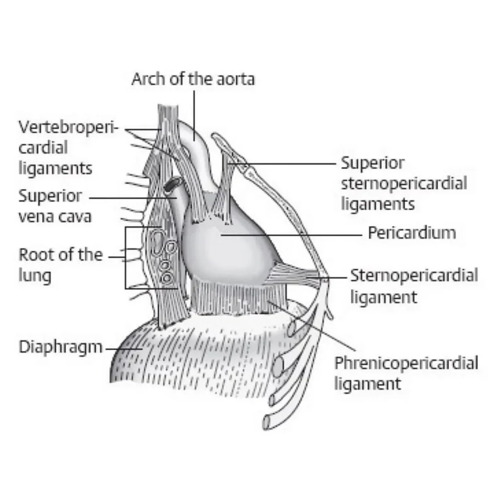
When the heart muscle arteries are tight, they pull on the ligaments that keep the heart correctly positioned in the thoracic cavity.
These heart ligaments are:
- Vertebropericardial ligaments, connecting the heart to vertebral bodies from C4 to T4
- Superior and inferior sternopericardial ligaments, connecting the heart to the sternum
- Phrenicopericardial ligaments, connecting the heart to the diaphragm
Here’s my favorite example:
Gentle traction of the carotid arteries with intention into the heart causes relaxation of arteries in the heart muscle.
When the heart muscle arteries are tight, they pull on the ligaments that keep the heart correctly positioned in the thoracic cavity.
These heart ligaments are:
- Vertebropericardial ligaments, which connect the heart to vertebral bodies from C4 to T4.
- Superior and inferior sternopericardial ligament, which connects the heart to the sternum.
- Phrenicopericardial ligaments, which connect the heart to the diaphragm.
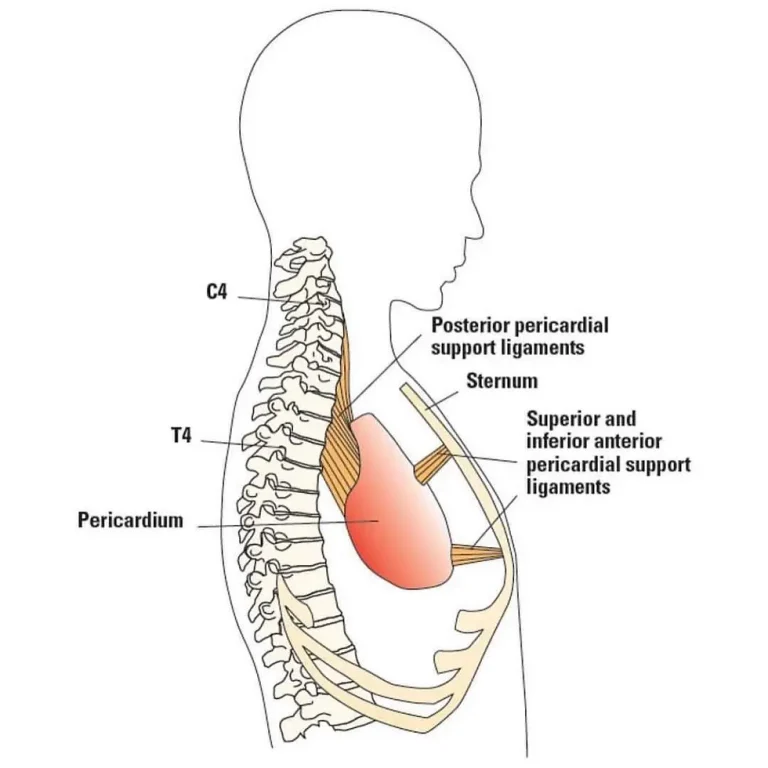
When the arterial vessels in the heart relax, all of these connective tissue connections soften, causing relaxation of the cervical and thoracic spines, as well as the diaphragm and sternum. This release of spinal tension thereby straightens the crooked spine. This includes relaxation of ribs 6 through 12, which are connected to the diaphragm that is attached to the heart via the phrenicopericardial ligament.
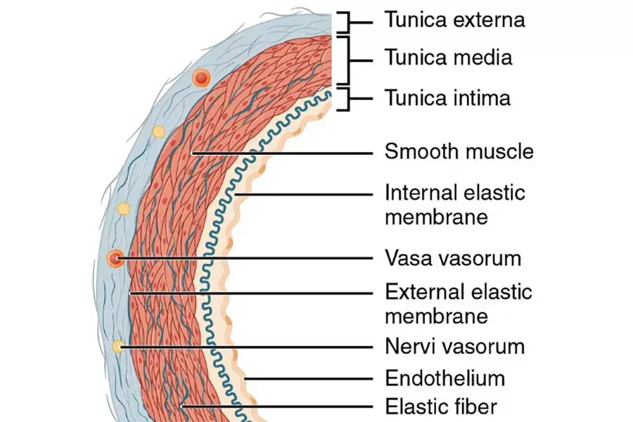
When the practitioner gently pulls on an artery, the artery gently pulls back.
Arteries have elastic and muscular layers. When the practitioner pulls on the restricted vessel(s), they pull back.
The practitioner lets them win the “Tug-of-War.”
The practitioner pulls on the arteries and the arteries pull back. Then the arteries relax, and blood flow increases.
This “Tug-of-War” only lasts a few seconds.
In the end, there is complete relaxation without any resistance noticed in the arteries in the heart, and you may palpate a significant increase in blood flow.
We body workers have been treating musculoskeletal symptoms through bodywork rather than addressing the primary cause, which is arterial constrictions.
Commonly, tension in the spine is secondary to tension in the vascular system.
HOW TO SCREEN FOR SCOLIOSIS
In learning how to correct scoliosis, we must first find it. Screening for scoliosis is a science—it is methodical, standardized, and repeatable.
Scoliosis screening for one spine takes less than a minute.
I subjectively grade scoliosis from mild-mild to severe-severe:
- Mild-Mild
- Mild-Moderate
- Mild-Severe
- Moderate-Mild
- Moderate-Moderate
- Moderate-Severe
- Severe-Mild
- Severe-Moderate
- Severe-Severe
For my video and book, which include spinal screening for scoliosis, go to my website: www.QuickSelfFixes.com .
Next, go to “Free Videos & Books” and scroll down to “Scoliosis Resolution.”
In the book, the chapter “How to Screen for Scoliosis” is on page two. In the video, this chapter begins at two minutes into the video.
Please learn how to screen for scoliosis, and teach a friend how to screen for scoliosis so they may screen you for scoliosis.
Help us help people heal from scoliosis and the discomfort it causes.
Dr. Cam & the Practitioners He Trains — And the Results They’re Achieving
Dr. Cassius Clay, Chiropractor
-
Case 14 - Pre Treatment - 1/06/26
-
Case 14 - Post Treatment
-
Case Study 1 - Pre Treatment -9/15/25
-
Case Study 1 - Post Treatment
-
Case Study 2 - Pre Treatment - 9/27/25
-
Case Study 2 - Post Treatment
-
Case Study -3 - Pre Treatment - 11/1/25
-
Case Study 3 - Post Treatment
-
Case Study 4 - Pre Treatment - 12/8/25
-
Case Study 4 - Post Treatment
-
Case Study 5 - Pre Treatment - 12/19/25
-
Case Study 5 - Post Treatment
-
Case Study 6 - Pre Treatment - 12/16/25
-
Case Study 6 - Post Treatment
-
Case Study 7 - Pre Treatment - 12/15/25
-
Case Study 7 - Post Treatment
-
Case Study 8 - Pre Treatment - 12/3/25
-
Case Study 8 - Post Treatment
-
Case Study 9 - Pre Treatment - 11/16/25
-
Case Study 9 - Post Treatment
-
Case Study 10 - Pre Treatment - 11/16/25
-
Case Study 10 - Post Treatment
-
Case Study 11 - Pre Treatment - 11/16/25
-
Case Study 11 - Post Treatment
-
Case Study 12 - Pre Treatment - 12/21/25
-
Case Study 12 - Post Treatment
-
Case Study 13 - Pre Treatment - 9/6/25
-
Case Study 13 - Post Treatment
-
Case 14 - Pre Treatment - 1/06/26 
-
Case 14 - Post Treatment -
Case Study 1 - Pre Treatment -9/15/25 -
Case Study 1 - Post Treatment -
Case Study 2 - Pre Treatment - 9/27/25 -
Case Study 2 - Post Treatment -
Case Study -3 - Pre Treatment - 11/1/25 -
Case Study 3 - Post Treatment -
Case Study 4 - Pre Treatment - 12/8/25 -
Case Study 4 - Post Treatment -
Case Study 5 - Pre Treatment - 12/19/25 -
Case Study 5 - Post Treatment -
Case Study 6 - Pre Treatment - 12/16/25 -
Case Study 6 - Post Treatment -
Case Study 7 - Pre Treatment - 12/15/25 -
Case Study 7 - Post Treatment -
Case Study 8 - Pre Treatment - 12/3/25 -
Case Study 8 - Post Treatment -
Case Study 9 - Pre Treatment - 11/16/25 -
Case Study 9 - Post Treatment -
Case Study 10 - Pre Treatment - 11/16/25 -
Case Study 10 - Post Treatment -
Case Study 11 - Pre Treatment - 11/16/25 -
Case Study 11 - Post Treatment -
Case Study 12 - Pre Treatment - 12/21/25 -
Case Study 12 - Post Treatment -
Case Study 13 - Pre Treatment - 9/6/25 -
Case Study 13 - Post Treatment
Dr. Carl Amodio, Chiropractor
-
Case Study 1 - Post Treatment
-
Case Study 1 - Pre Treatment -10/12/25
-
Case Study 2 - Pre Treatment -10/29/25
-
Case Study 2 - Post Treatment
-
Case Study 1 - Post Treatment -
Case Study 1 - Pre Treatment -10/12/25 -
Case Study 2 - Pre Treatment -10/29/25 -
Case Study 2 - Post Treatment